编者按:慢性完全闭塞病变(CTO)一直是PCI介入术者面临的难题和挑战,复杂高危病例的治疗更是难点。首都医科大学附属北京安贞医院宋现涛教授介绍了一例复杂双支CTO病变PCI术式。
病例资料
主诉 男性,45岁。因“劳力性胸痛3个月,加重2天”入院。
现病史 2型糖尿病、高血压病、高脂血症。
体格检查 心电图无异常。甘油三酯(TG)2.09 mmol/L,总胆固醇(TC)6.04 mmol/L,低密度脂蛋白胆固醇(LDL-C)4.29 mmol/L,肌钙蛋白阴性。尿素氮6.3 mmol/L,Cr93.9 μmol/L。无明确阳性体征。
辅助检查 冠状动脉造影示右冠状动脉(RCA)全程多发斑块,局部闭塞,左冠状动脉近端偏心性斑块,局部重度狭窄。
入院诊断 不稳定性心绞痛、2型糖尿病、高血压病3级(极高危)、高脂血症
治疗方案
1. 双联抗血小板、降糖、扩冠、调脂;
2. 择期冠脉造影(图1、图2);
3. 对比剂(威视派克)
(1).gif)
.gif)
图1. 冠状动脉造影
.gif)
图2. 右冠状动脉造影
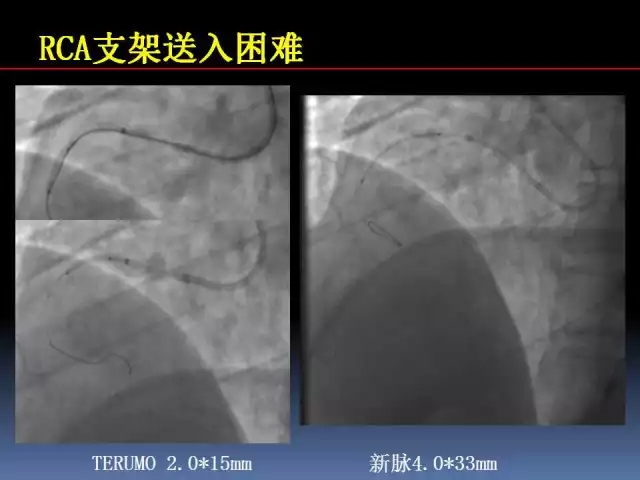
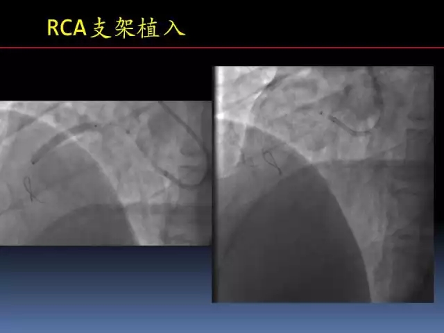
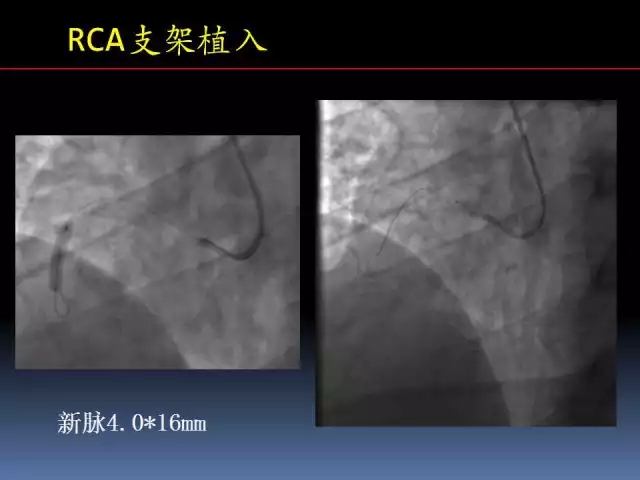
建议首选冠状动脉旁路移植术(CABG)。患者较年轻,家属要求尝试经皮冠状动脉介入治疗(PCI)。先处理右冠冠状动脉,选桡动脉途径入路(图3)。选择强支撑力的指引导管。
.gif)
图3. 开通右冠状动脉后
.jpg)
一周后行左冠状动脉慢性完全闭塞病变介入治疗。策略为经股动脉左冠状动脉PCI,经桡动脉对侧造影(图4~8)。
.gif)
.gif)
图4. 复查右冠脉造影
.gif)
.gif)
图5. 对侧造影指导导丝通过闭塞病变
.gif)
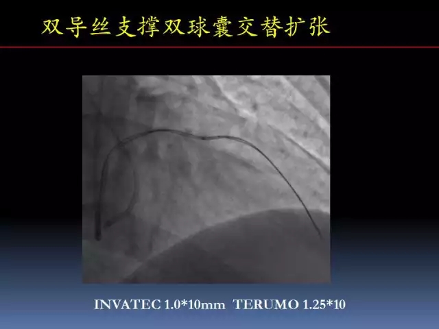
图6. 球囊不能通过病变
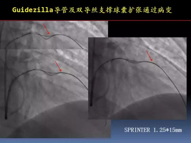
.gif)
图7. LAD球囊扩张及支架定位

图8. LAD支架释放后造影
术后处理
低分子肝素抗凝治疗3天。血小板聚集率9%,血小板最大聚集率11%。择期处理回旋支病变。
.jpg)